Scanning Electron Microscopy / Energy Dispersive Spectroscopy
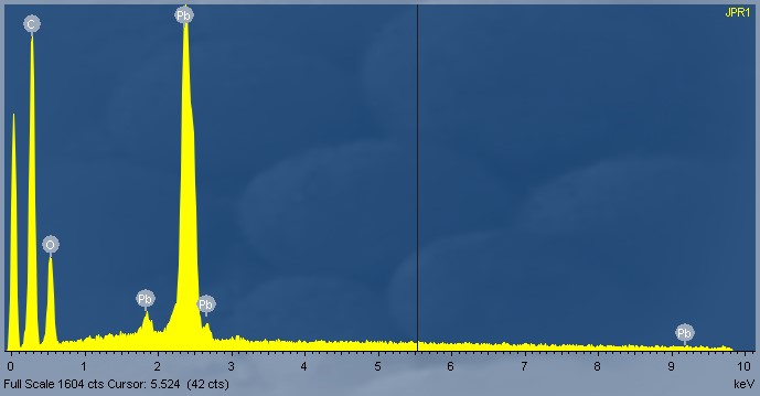
SEM-EDS analysis is a non-destructive analytical technique (to the sample), but unlike XRF that can be undertaken in-situ without sample removal, SEM-EDS does require sample removal. The sample material is irradiated with electrons resulting in the emission of x-rays characteristic to the elements present. The energy emissions are translated into spectral peaks of varying intensity, resulting in a spectrum profile (see image below), which identifies the different inorganic elements present in the sample (ie lead, iron, copper, zinc etc.).
The analysis is not quantitative. The X-ray intensity (size of spectrum peaks) is directly proportional to the concentration of the elements in the sample. This technique is able to characterise individual paint layers or individual particles within a sample, making the analysis very targeted and specific to particular areas of interest (ie such as individual layers of a cross-section).
The results are interpreted and presented in a report, with the EDS spectra included (see Image 1 below).
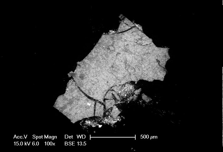
The high powered Scanning Electron Microscope enables examination of small sample particles in high resolution, up to 50, 000 x (see Image 2 below). This enables assessment of the surface of a sample, individual components of a sample and targeted analysis of the sample.